

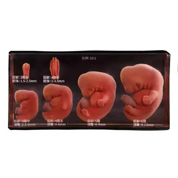
Medical curricula face a persistent pedagogical bottleneck when introducing early human development. Students must conceptualize highly dynamic, microscopic transformations across three dimensions within a tightly compressed developmental timeline. Flat illustrations in textbooks fail to convey these changes, forcing learners to mentally reconstruct complex structural folding and tissue migrations.
To bridge this spatial gap, modern medical universities are upgrading their laboratory environments with advanced physical models. By translating micro-scale biological landmarks into high-fidelity physical assets, institutions can now provide clinical students with clear, hands-on spatial frameworks that simplify complex anatomical pathways.
The Visual Limitations of Two-Dimensional Curriculum Materials
During embryogenesis, structures like the neural tube, pharyngeal arches, and somites form concurrently in a highly confined space. Correctly visualizing how the ectoderm, mesoderm, and endoderm orient themselves relative to one another requires outstanding spatial awareness. Standard line diagrams and static slides fail to convey the true depth and curvature of these early formations.
When students try to memorize these structures purely from flat screens or printed pages, they often struggle with spatial orientation during clinical rotations and imaging interpretation. This lack of clear spatial understanding directly impacts their ability to recognize anatomical relationships in three-dimensional environments. The disconnect highlights the critical need for a practical, tactile solution in modern anatomical classrooms.
Transforming High-Resolution Digitized Volumetric Data into Physical Reality
Addressing this instructional challenge requires a level of manufacturing accuracy that matches actual human biology. Standard commercial teaching aids often simplify or distort complex pathways, diminishing their clinical utility. DIGIHUMAN resolves this issue by extracting anatomical specifications directly from ultra-high-resolution digitized human data sets.
The foundational dataset features a precise voxel size of 0.0384mm by 0.0384mm by 0.1mm. High-precision segmentation systems map this data directly, ensuring every minor structure is represented with high anatomical accuracy. To improve anatomical accuracy, formalin-fixed human specimens serve as the direct reference throughout the modeling phase. This rigorous comparison process ensures that the physical replica provides a highly accurate visual and spatial experience that mimics actual human anatomy.
Multi-Channel Jetting and Material Specifications
Accurately representing early developmental tissues requires advanced hardware capabilities. Traditional single-material 3D printing cannot differentiate the distinct layers, variable densities, and subtle color gradients of a developing embryological structure. DIGIHUMAN addresses this by employing advanced 3D inkjet printing and light curing technology.
The printing system features 12 independent material channels, allowing for the simultaneous deposition and light-curing of diverse resins formulas. This multi-material capacity enables unique printing combinations, such as single hardness sections for dense structures and soft-and-hard combinations for realistic tissue transitions. Furthermore, crystal-clear transparent outer resins can enclose delicate inner pathways. This transparent packaging technique allows students to observe the exact positioning of internal organ precursors—such as the early vascular network or branchial structures— while keeping the entire structure visible and protected.
Integrating Physical Models into Laboratory Workflows
Bringing physical, high-simulation models into medical school laboratories transforms how students process spatial information. Tactile exploration engages muscle memory and accelerates anatomical recognition during active study sessions. For institutions looking to modernize their curricula, adopting a high-fidelity embryo 3d model manufactured by DIGIHUMAN provides a direct educational advantage.
These models scale microscopic developmental stages into easily observable, full-color macroscopic teaching aids. Professors can effortlessly demonstrate how the primitive gut tube folds, how the pericardial cavity positions itself, or how early vascular networks progress across sequential stages. Students can rotate the physical unit to trace complex developmental paths from any angle. This direct, hands-on engagement reduces the frustration of mentally translating flat textbook illustrations into three dimensions, supporting improved knowledge retention.
Maximizing Institutional Resource and Laboratory Efficiency
Upgrading to high-fidelity anatomical models provides significant long-term administrative value for university purchasing departments. Preserving real biological specimens involves high recurring costs, specialized ventilation systems, strict hazardous material compliance, and extensive maintenance protocols. Furthermore, rare developmental specimens are fragile, difficult to acquire ethically, and naturally degrade over time under heavy student handling.
Reconstructed physical models made from environmentally friendly, durable resins reduce many of these compliance requirements associated with biological specimens. Students can handle these models continuously during self-directed study sessions without risking damage to delicate structures. This physical durability guarantees a consistent, highly repeatable educational standard across successive student cohorts. This allows universities to reduce annual laboratory overhead while offering premium training resources.
Synchronizing Tangible Assets with Digital Dissection Systems
Physical learning tools deliver the best educational outcomes when paired with interactive digital systems. Medical universities can create complete learning ecosystems by combining these detailed models with digital platforms like the DIGIHUMAN Virtual Anatomy Table.
Under this approach, students first perform virtual dissections on high-definition 4K multi-touch screens, tracing structural networks, toggling layers, and isolating microscopic tissues. They then move to the physical model to gain the tactile feedback that digital screens alone cannot provide. This integrated learning loop connects digital clarity with physical handling, accelerating spatial comprehension and preparing future physicians for clinical success.
